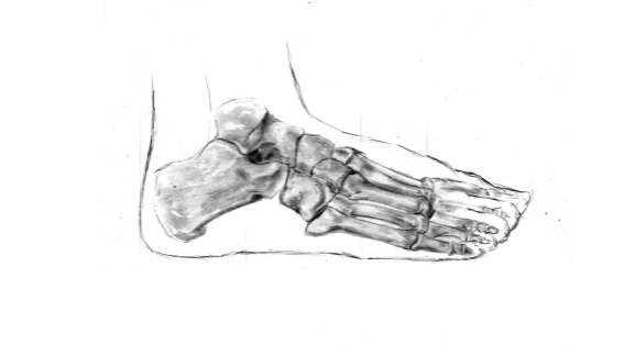
Foot Skeletal Anatomy
Overview
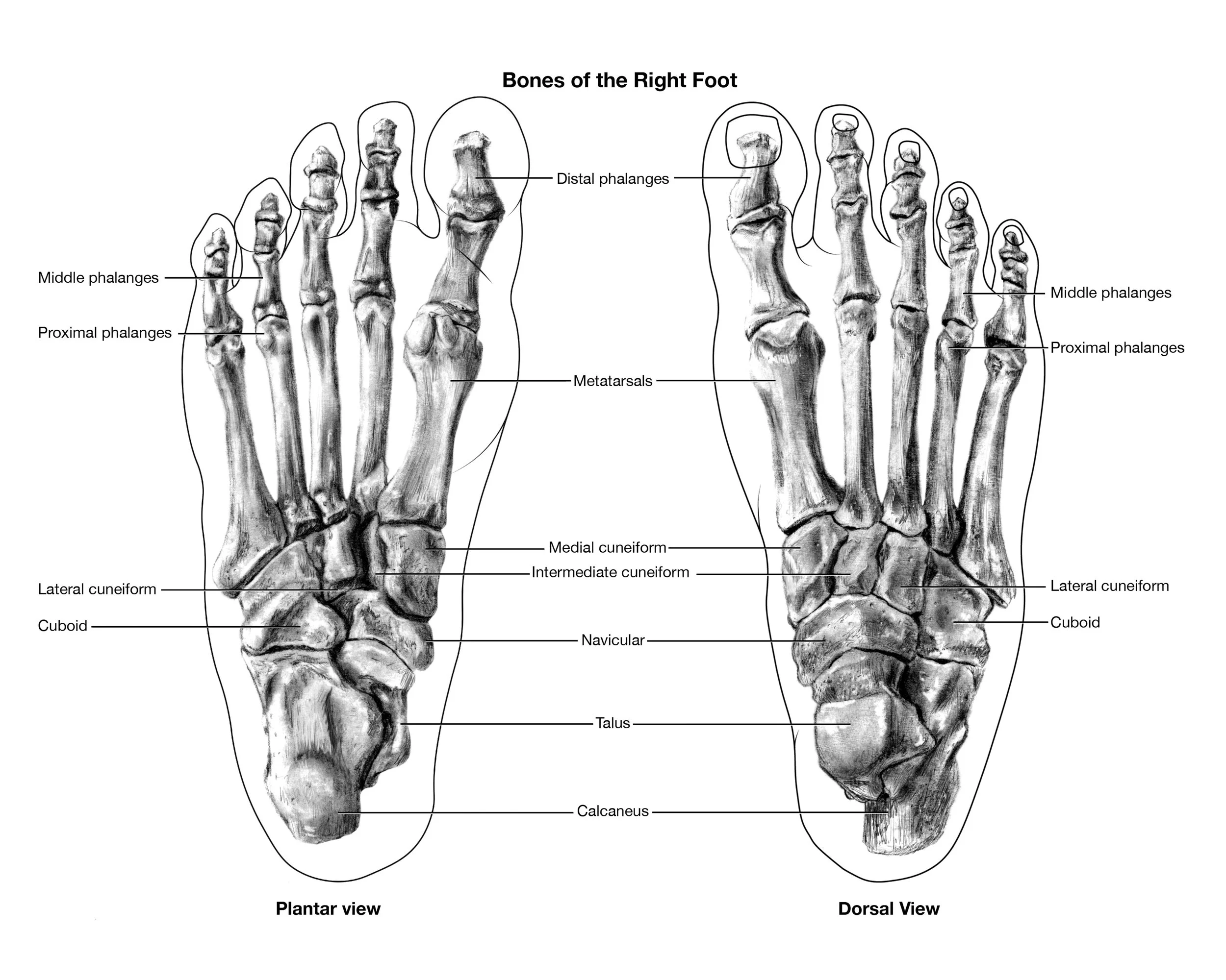
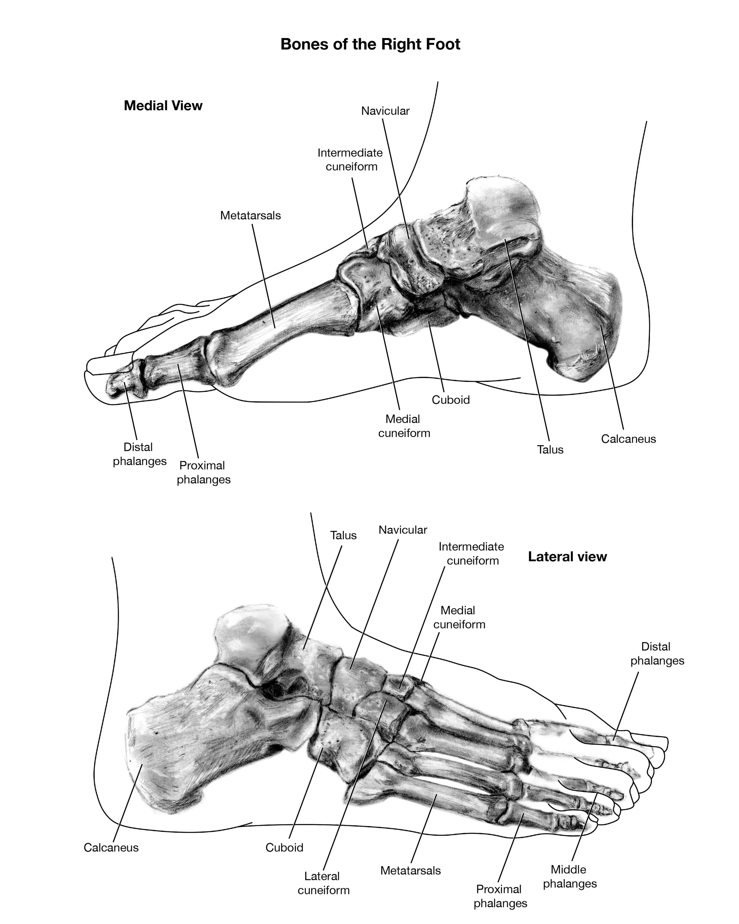
A study of human foot anatomy designed to communicate skeletal structure through clear educational visualization. The project focuses on anatomical accuracy, spatial relationships, and instructional clarity.
Process
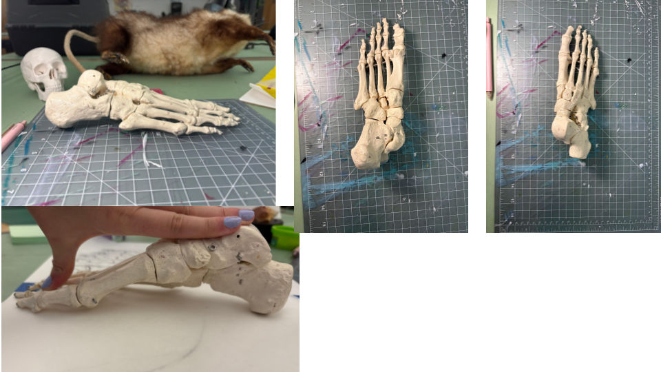
Anatomical references and observational studies were used to establish accurate proportions and identify major skeletal landmarks. Multiple views were developed to support spatial understanding.
Final Outcome
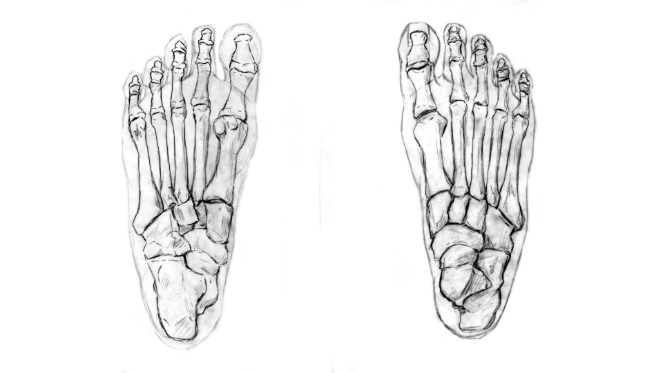
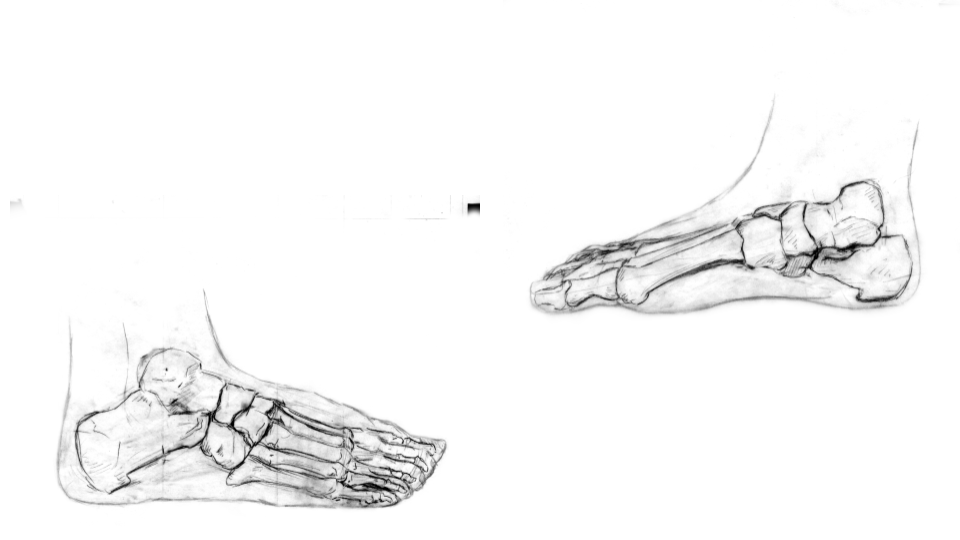
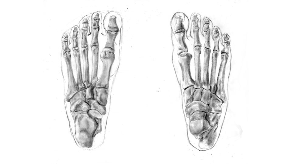
The final illustrations present the skeletal anatomy of the foot from several perspectives, creating an accessible educational reference.
Skills
Medical Illustration • Human Anatomy • Scientific Visualization • Educational Design