
Polycystic Ovary Syndrome (PCOS): Anatomy & Pathophysiology
Medical Illustration • Science Communication • Information Design
Project Overview
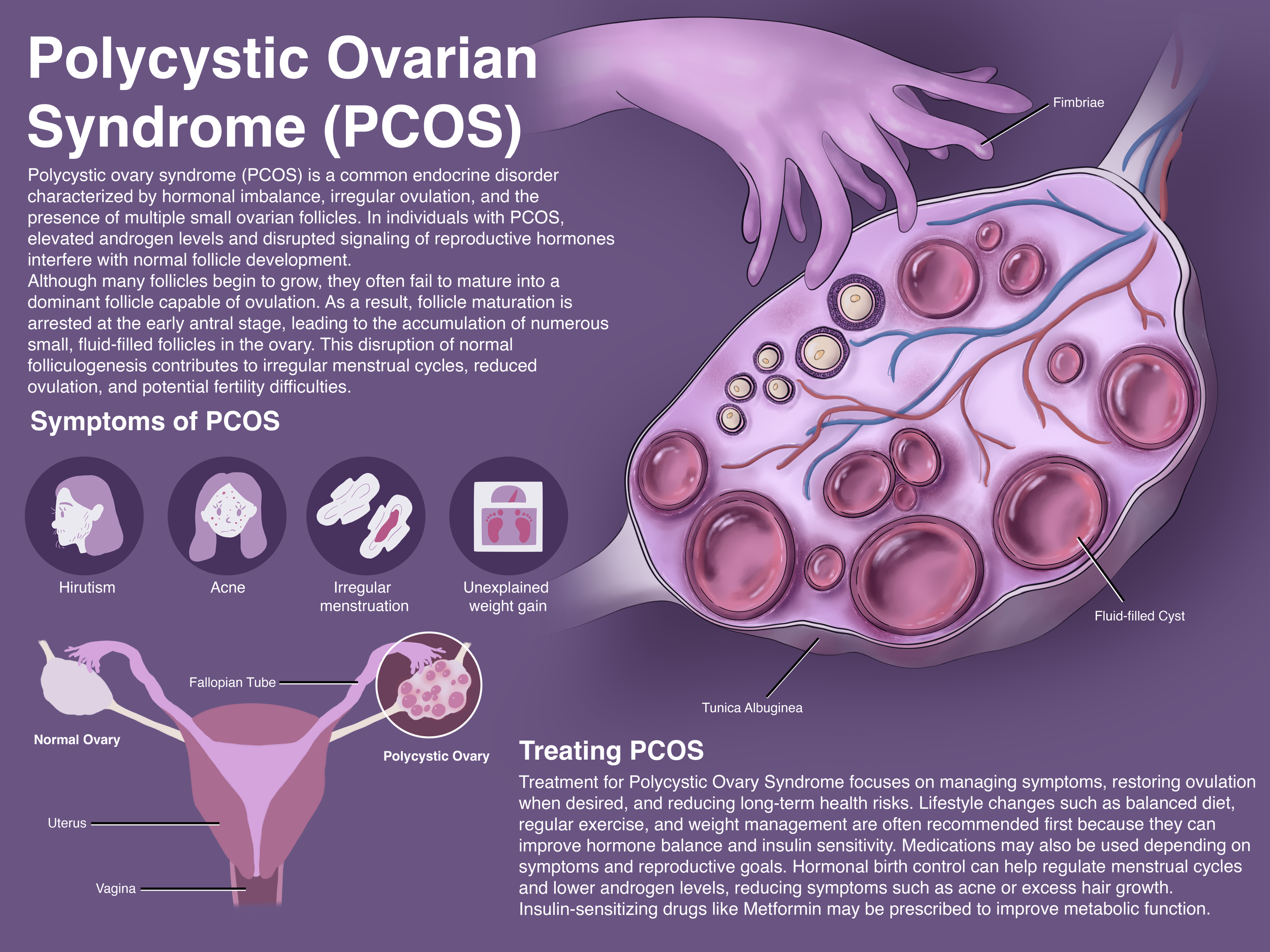
This project explores how medical illustration can be used to communicate reproductive health concepts to non-specialist audiences. Beginning with an educational cross-section of a healthy ovary, the project evolved into a companion piece illustrating the anatomical and physiological changes associated with Polycystic Ovary Syndrome (PCOS).
The final poster combines anatomical illustration, scientific writing, and information design to explain how hormonal dysregulation affects follicle development and ovarian function. The goal was to create an educational resource that balances scientific accuracy with accessibility, making a complex endocrine disorder easier to understand.
The Challenge
Polycystic Ovary Syndrome is one of the most common endocrine disorders affecting reproductive-age individuals, yet many explanations rely heavily on medical terminology and dense text.
The objective of this project was to transform complex physiological processes into a visual format that could be understood by a broad audience. To accomplish this, I needed to accurately represent ovarian anatomy while explaining how altered hormone signaling disrupts normal follicle maturation and ovulation.
A secondary challenge was adapting an existing healthy ovarian cross-section into a pathology-focused educational illustration while maintaining anatomical consistency and scientific accuracy.
Research and Process
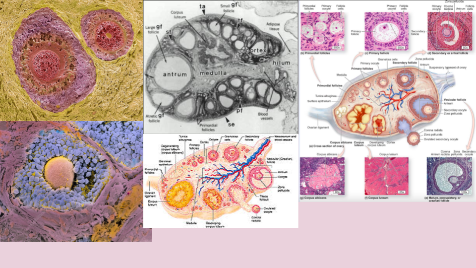
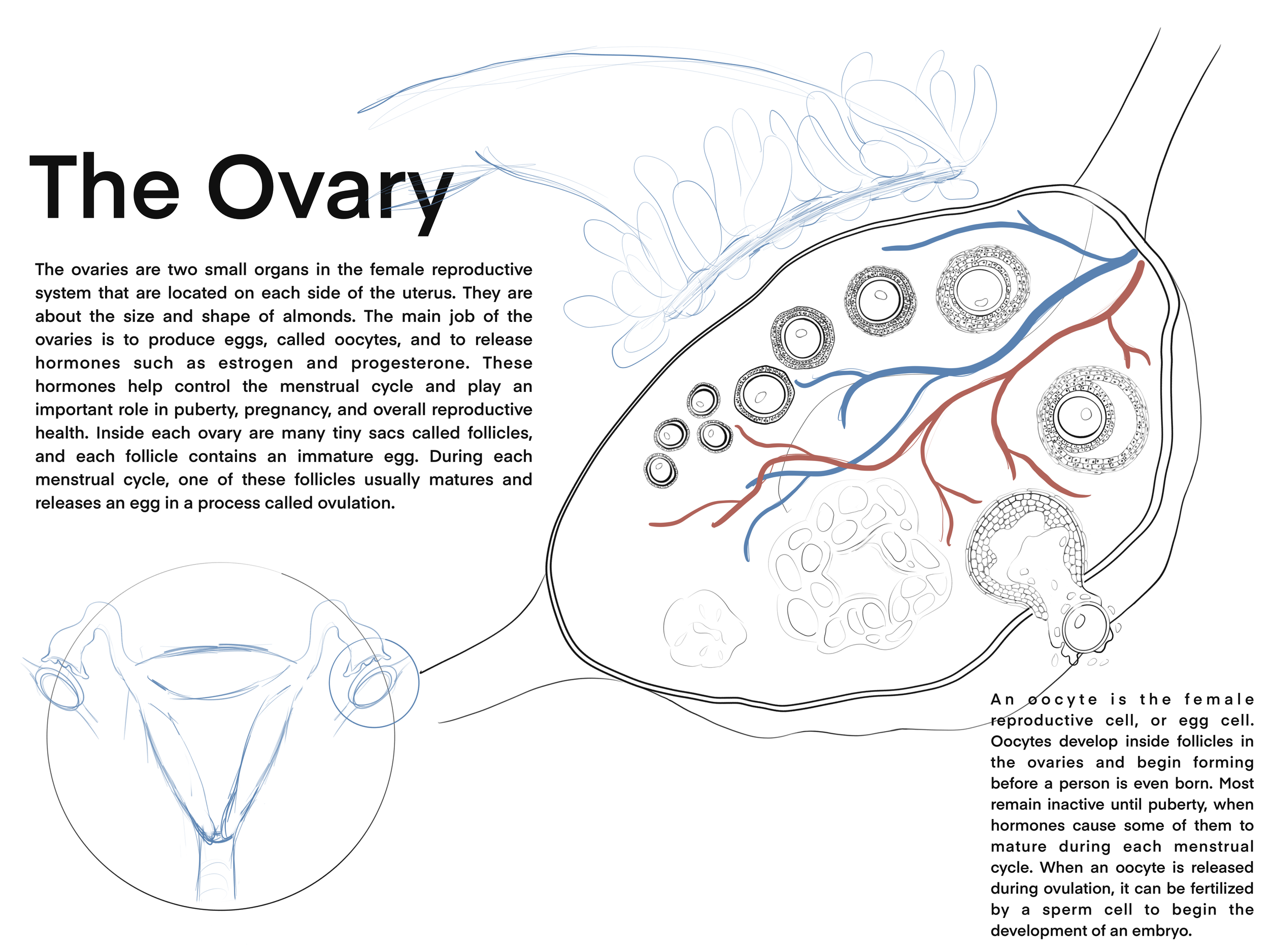
The project began with the development of a detailed ovarian cross-section illustrating normal follicular development. Research focused on ovarian histology, folliculogenesis, and reproductive physiology to ensure anatomical accuracy. The initial poster visualized the progression from primordial follicles to ovulation and the formation of the corpus luteum.
After establishing the healthy anatomy, the project expanded to investigate the physiological mechanisms underlying PCOS. Research included endocrine regulation, androgen excess, follicular arrest, ovulatory dysfunction, and current treatment approaches. Particular attention was given to how hormonal imbalance prevents the maturation of a dominant follicle, resulting in the accumulation of numerous small follicles within the ovary.
The visual design process involved:
Anatomical reference gathering
Histological and physiological research
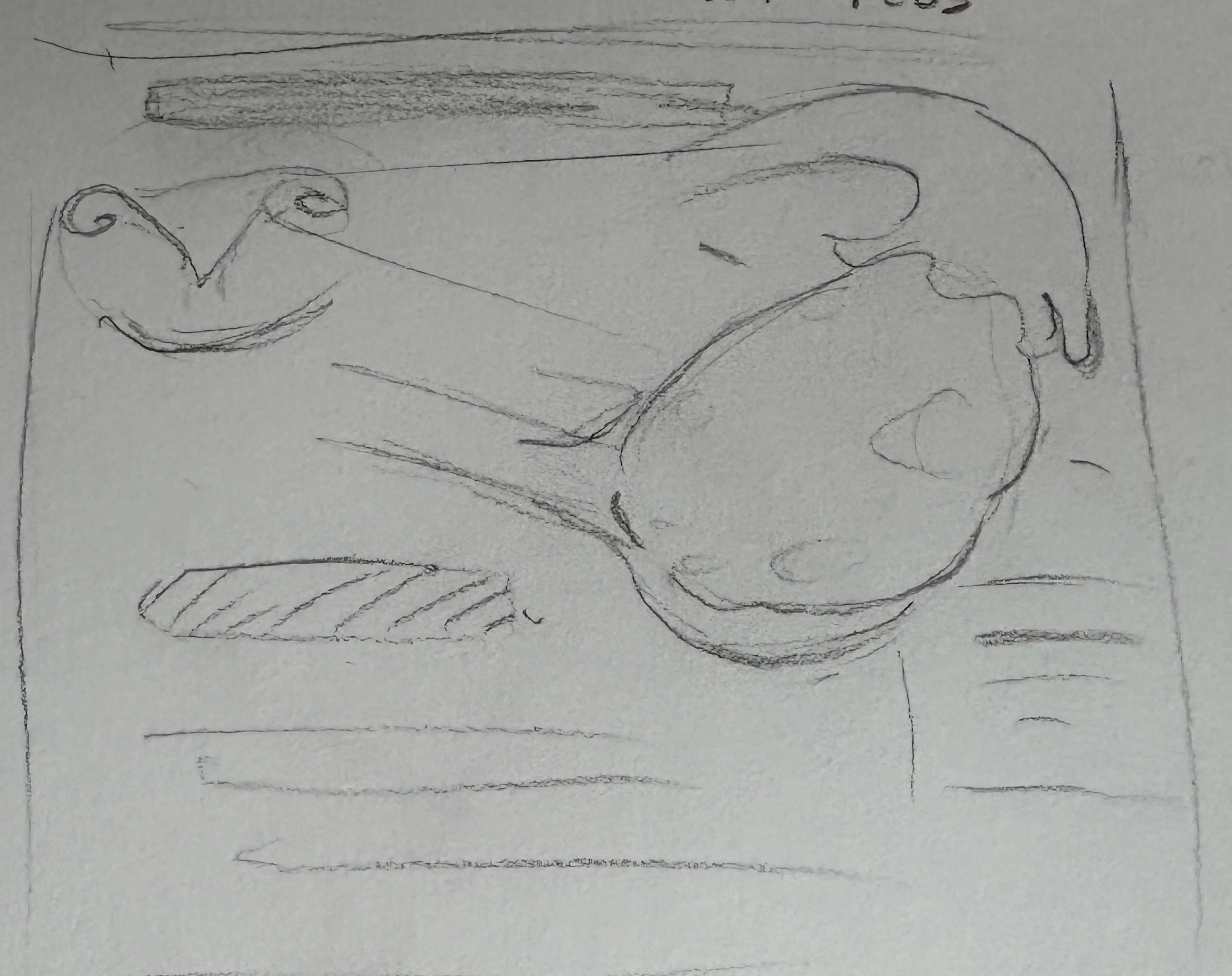
Cross-sectional anatomical drawing
Content organization and educational writing
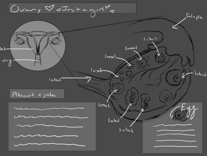
Information hierarchy development
Layout refinement and visual integration
Throughout the project, scientific accuracy remained the primary consideration while ensuring the information remained approachable for educational audiences.
Final Outcomes
The final deliverable is an educational medical illustration comparing normal ovarian anatomy with the structural and physiological changes associated with PCOS.
Educational Ovarian Cross Section
The first phase of the project focused on creating an educational illustration of a healthy human ovary. The objective was to accurately depict ovarian anatomy while explaining the process of follicular maturation and ovulation. The final poster visualizes the progression of follicles from primordial follicles through the Graafian follicle stage, as well as the formation of the corpus luteum and corpus albicans following ovulation.
Special attention was given to anatomical accuracy, tissue organization, and educational clarity. Labels, explanatory text, and visual hierarchy were incorporated to support learning and provide context for the physiological processes occurring within the ovary. The completed illustration serves as an educational resource introducing normal ovarian structure and reproductive function.
Together, the series transforms scientific research into approachable educational materials that support learning, engagement, and public understanding.
Polycystic Ovarian Syndrome (PCOS) Educational Poster
Building upon the healthy ovarian cross section, the second phase of the project adapted the illustration to communicate the pathological changes associated with Polycystic Ovary Syndrome (PCOS). Research focused on endocrine regulation, follicular arrest, androgen excess, and the disruption of normal ovulatory cycles.
The final poster combines anatomical illustration and educational content to explain how altered hormone signaling interferes with follicle maturation. Rather than progressing toward ovulation, follicles become arrested during development and accumulate within the ovary, contributing to many of the reproductive symptoms associated with PCOS.
To support audience understanding, the poster also includes concise explanations of the condition, its physiological effects, and common treatment approaches. The result is an educational resource that transforms complex endocrine concepts into an accessible visual narrative while maintaining scientific accuracy.
Reflection
This project strengthened my ability to synthesize information from anatomy, physiology, developmental biology, and endocrinology into a cohesive visual narrative. It reinforced the importance of medical illustration as a communication tool capable of supporting health literacy and public understanding of complex biological systems.
Skills Utilized
• Medical Illustration
• Scientific Communication
• Information Design
• Anatomy
• Endocrine Physiology
• Research Synthesis
• Educational Writing
• Adobe Illustrator
• Adobe Photoshop
• Visual Storytelling
• Scientific Visualization